
In the dynamic landscape of modern medicine, keeping pace with rapid advancements isn’t just a goal; it’s a professional imperative. For specialists in diagnostic and therapeutic procedures, the field of interventional radiology (IR) stands out, continually evolving with new techniques that redefine patient care. These innovations demand a commitment to lifelong learning, ensuring clinicians can leverage the most effective, minimally invasive approaches available.
We understand the demands placed on healthcare professionals to remain at the forefront of their specialties. The rapid integration of sophisticated imaging, novel devices, and procedural refinements means today’s practitioners must constantly update their knowledge to maintain clinical excellence. Our commitment at Edu Symp, as a premier continuing medical education provider operating since 1975, is to deliver quality and accredited learning opportunities that empower you to do just that.
What Is Interventional Radiology? A Foundation
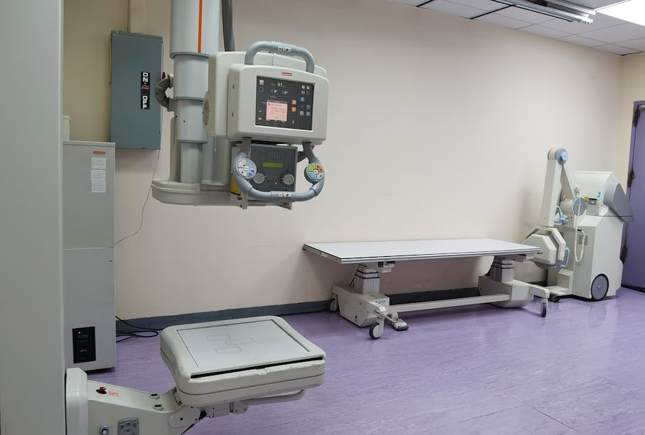
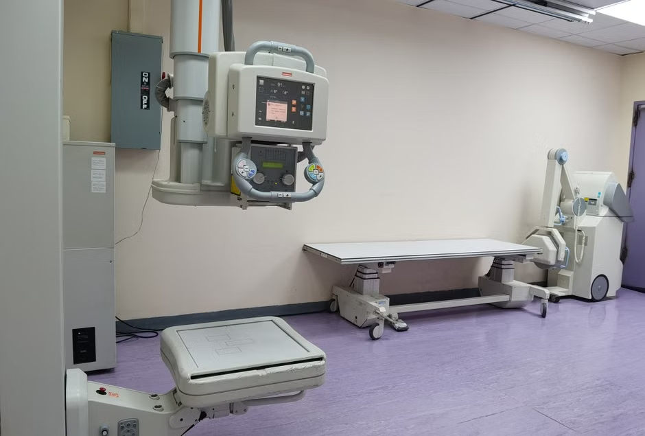
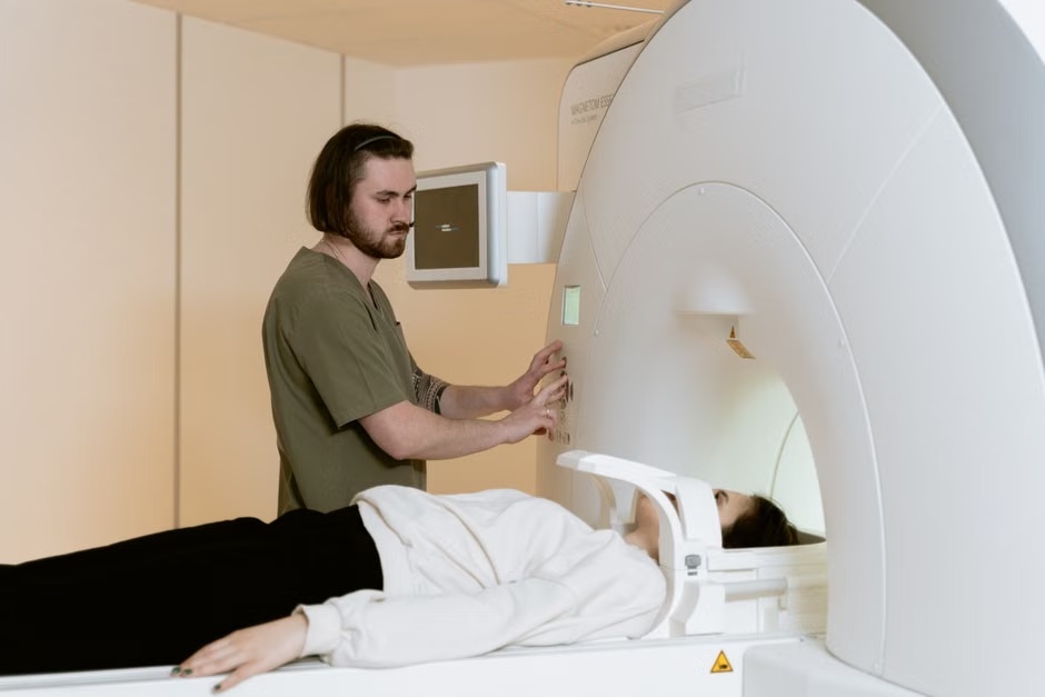
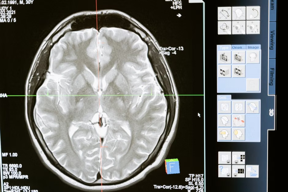
Interventional radiology is a medical subspecialty that uses minimally invasive, image-guided procedures to diagnose and treat diseases in nearly every organ system. Rather than traditional open surgery, interventional radiologists employ advanced imaging technologies—such as X-rays, CT, ultrasound, and MRI—to guide tiny instruments, like catheters and wires, through the body. This approach often leads to less pain, smaller incisions, and faster recovery times for patients compared to conventional surgical methods.
The core principle is precision. Every intervention, from biopsies to tumor ablations, is performed under real-time imaging, ensuring accuracy and minimizing risk. As defined by the Johns Hopkins Medicine Health Library, interventional radiology offers an array of treatments for a wide variety of conditions without surgery.
How Do Interventional Radiology Procedures Work?
Interventional radiology procedures typically begin with a detailed imaging study to map the target area. Once the precise location is identified, a small incision, often just a few millimeters, is made. Guided by live imaging, a thin catheter or needle is carefully inserted through this incision and navigated to the treatment site. Contrast agents may be used to enhance visibility of blood vessels or organs. Once in place, various tools can be deployed through the catheter to perform the necessary intervention, whether it’s delivering medication, removing blockages, or treating tumors.
This image-guided methodology is what differentiates IR. It enables highly targeted treatments while minimizing trauma to surrounding healthy tissues. For example, in our practice, we’ve seen how effectively image guidance can reduce procedural complications, especially when dealing with complex vascular structures or deep-seated lesions. The ability to visualize the anatomy in real-time is paramount for successful outcomes.
What Conditions Can Interventional Radiology Treat?
Interventional radiology has broad applications across numerous medical conditions. Its minimally invasive nature makes it suitable for patients who may not be candidates for open surgery, or those seeking quicker recovery. From vascular diseases to oncology, the scope continues to expand. We equip our learners with the knowledge to apply these techniques in their daily practice.
Some of the conditions commonly treated by interventional radiologists include:
- Vascular Diseases: Peripheral artery disease, deep vein thrombosis (DVT), varicose veins, aneurysms, and embolisms.
- Oncology: Liver, kidney, and lung tumor ablation; chemoembolization and radioembolization for cancer treatment; biopsy for diagnosis.
- Women’s Health: Uterine fibroid embolization (UFE), pelvic congestion syndrome treatment.
- Men’s Health: Prostate artery embolization (PAE) for benign prostatic hyperplasia.
- Gastrointestinal Issues: Biliary obstructions, liver disease (e.g., TIPS procedures), feeding tube placements.
- Kidney Disease: Renal artery stenosis, dialysis access maintenance.
- Pain Management: Vertebroplasty and kyphoplasty for spinal fractures, nerve blocks.
Recent Advances in Interventional Radiology
The field of interventional radiology is marked by continuous evolution, driven by technological innovations and a deeper understanding of disease processes. These recent advances are not just incremental; they represent significant leaps forward in precision, safety, and patient outcomes. From refined imaging techniques to novel therapeutic agents, the landscape of IR is always shifting, presenting new opportunities for patient care.
We’re observing substantial progress in areas like advanced imaging fusion, which combines real-time ultrasound with pre-acquired CT or MRI data for unparalleled guidance during complex procedures. There’s also significant growth in interventional oncology, with more targeted delivery methods for chemotherapy and radiation, minimizing systemic side effects. Such developments enhance treatment efficacy and expand the applicability of IR to a broader patient population.
“Interventional radiology is at the forefront of medical innovation, continually developing less invasive ways to treat conditions that once required major surgery. The emphasis on image guidance ensures both precision and safety, fundamentally changing how we approach patient care.”
AI in Interventional Radiology: Current Concepts and Future Trends
Artificial intelligence (AI) is rapidly transforming interventional radiology by enhancing image analysis, improving procedural planning, and optimizing workflow. AI algorithms can detect subtle anomalies in imaging studies, predict treatment responses, and assist in real-time guidance during interventions, thereby increasing accuracy and efficiency for radiology professionals. This integration promises to elevate diagnostic precision and therapeutic effectiveness.
Current applications of AI in interventional radiology focus on several key areas. Machine learning models are being developed to segment tumors more precisely from healthy tissue, aiding in pre-procedural planning for ablation therapies. We’re seeing AI used to automate tedious measurement tasks in vascular imaging, potentially reducing procedural time. Furthermore, AI can help predict patient outcomes, allowing for more personalized treatment strategies. The future trends suggest an even deeper integration, with AI assistants providing cognitive support during complex cases and automating image interpretation in high-volume settings.
In our neuroradiology programs, we often discuss how AI could revolutionize the identification of subtle abnormalities and assist in guiding intricate procedures, particularly in areas requiring extreme precision. This could significantly impact the practice of neuroradiology in clinical practice.
Robotics in Interventional Radiology
Robotics are increasingly integrated into interventional radiology to enhance precision, stability, and control during complex procedures. Robotic systems can perform intricate tasks with sub-millimeter accuracy, surpass human dexterity, and minimize radiation exposure for operators by allowing remote control. This technology is particularly beneficial for highly repetitive or extremely precise interventions, pushing the boundaries of what’s surgically possible with minimal invasiveness.
The use of robotics in interventional radiology is evolving from simple needle guidance systems to sophisticated platforms capable of autonomous navigation and tool manipulation. These systems offer unparalleled stability, crucial for long, delicate procedures where even a slight tremor can have significant consequences. We’ve seen how robotic assistance can reduce operator fatigue and improve ergonomics, contributing to safer and more consistent outcomes. Beyond current applications, the development of haptic feedback systems and collaborative robots that work alongside the interventional radiologist is underway, promising to make complex procedures even more accessible and precise. The ability to perform delicate maneuvers with augmented precision, much like in surgical pathology updates, is becoming a hallmark of modern medical interventions.
“Robotics in interventional radiology are not replacing the physician, but augmenting their capabilities, allowing for unprecedented precision and enabling procedures that were once deemed too complex or risky. This represents a significant step forward in patient safety and clinical outcomes.”
Suitability: When Is Interventional Radiology the Right Choice?
Interventional radiology isn’t always the first or only option, but it offers distinct advantages in many scenarios. It’s often the right choice when patients require a minimally invasive approach due to comorbidities, age, or a desire for faster recovery. It’s also preferred for procedures that benefit from precise image guidance, such as targeted biopsies, localized drug delivery, or embolization of specific vessels.
However, there are situations where traditional open surgery or medical management may be more appropriate. Large, complex tumors requiring extensive resection, conditions needing significant anatomical reconstruction, or diffuse diseases that don’t lend themselves to localized treatment are examples. Our accredited programs feature discussions with international faculty leaders who explore these nuances, helping practitioners understand when to recommend IR versus other modalities. This balanced perspective is vital for optimal patient care.
What to Expect: Realistic Outcomes and Recovery
Patients undergoing interventional radiology procedures generally experience quicker recovery times compared to traditional surgery. Most procedures are outpatient or require only a short hospital stay. You’ll typically have a small incision, often covered with a bandage, and may experience some mild discomfort or bruising at the site. Pain management is usually straightforward, often managed with over-the-counter medication.
The timeline for full recovery varies significantly based on the procedure and individual patient health. For many minor interventions, patients can resume normal, light activities within a day or two. More complex procedures, like tumor ablations or significant embolizations, might require a week or more for full recovery. We emphasize realistic expectations, ensuring patients understand the process and potential recovery period. Our focus is always on achieving positive clinical outcomes with minimal patient disruption.
Practical Tips for Interventional Radiologists
Staying current and effective in a rapidly evolving field like interventional radiology requires dedication and strategic learning. Here are some practical tips we’ve cultivated over our decades of experience in medical education:
- Commit to Continuous Learning: Regularly attend accredited CME programs and symposia focused on advanced radiology techniques. The field changes quickly; staying updated is non-negotiable.
- Master Advanced Imaging: Develop deep expertise in all imaging modalities used for guidance (ultrasound, CT, MRI, fluoroscopy). Understanding their strengths and limitations is crucial.
- Embrace New Technologies: Be open to integrating AI tools and robotic assistance into your practice. Familiarize yourself with their operation and how they can enhance precision and efficiency.
- Network with Peers: Engage with other interventional radiologists at conferences and workshops. Sharing experiences and learning from complex cases can provide invaluable insights.
- Prioritize Patient Communication: Clearly explain procedures, potential risks, and expected outcomes to patients. Building trust and managing expectations is key to patient satisfaction.
- Focus on Subspecialty Expertise: While IR is broad, developing a niche (e.g., interventional oncology, vascular interventions) can allow for deeper expertise and more specialized referrals.
As interventional radiology continues its remarkable trajectory of innovation, the need for continuous, high-quality medical education becomes ever more apparent. At Edu Symp, we are committed to providing CME programs and symposia that not only cover the latest techniques and clinical applications but also integrate insights from international faculty leaders. Being an ACCME accredited provider, we guarantee that physicians experience trusted and high-quality educational experiences that will promote certification, clinical excellence, and lifelong learning. We believe that empowering healthcare professionals with practical and evidence-based knowledge is the pathway to advancing patient care across the nation. Join us in shaping the future of radiology.