
When a patient presents with sudden neurological deficits, the race against time begins. Every minute counts in acute stroke, and rapid, accurate neuroimaging is the cornerstone of effective management. Making the right decision between Computed Tomography (CT) and Magnetic Resonance Imaging (MRI) can profoundly impact patient outcomes, guiding critical interventions like thrombolysis or thrombectomy.
Physicians, radiologists, and other healthcare professionals understand the immense pressure involved in these moments. We need clear diagnostic pathways and a deep understanding of what each imaging modality offers. This isn’t just about identifying a stroke; it’s about characterizing its type, extent, and potential for intervention.
At Edu Symp, we’ve been an ACCME accredited provider since 1975, offering practical and evidence-based CME programs designed for real-world application. We understand the demands placed on healthcare professionals and are dedicated to supporting your continuous learning, especially in critical areas like stroke management. As we explain in our guide on stroke management: recognition, intervention, and rehabilitation, timely and appropriate imaging is paramount to preserving brain function.
Foundation: What is Acute Stroke and Why is Imaging Key?
An acute stroke occurs when blood flow to a part of the brain is interrupted, either by a blocked artery (ischemic stroke) or a ruptured blood vessel (hemorrhagic stroke). This interruption deprives brain cells of oxygen and nutrients, leading to rapid cell death. Early diagnosis and differentiation between ischemic and hemorrhagic stroke are crucial because treatment strategies differ significantly, and inappropriate treatment can cause severe harm. For instance, thrombolytic therapy, which dissolves clots, is life-saving for ischemic stroke but contraindicated in hemorrhagic stroke. Accurate imaging within the golden hour can dictate the entire course of treatment.
“Rapid neurological assessment and immediate neuroimaging are critical steps in the evaluation of acute stroke. Distinguishing between ischemic and hemorrhagic stroke is fundamental to guiding appropriate therapy and minimizing brain injury.”
— National Institute of Neurological Disorders and Stroke (NINDS)
MRI vs CT in Acute Stroke: The Initial Assessment
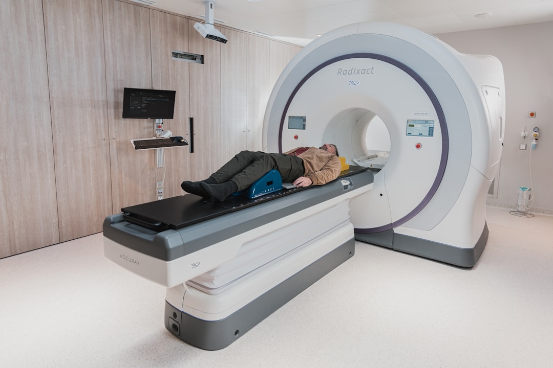
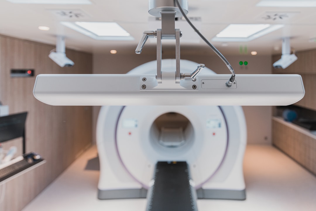
Computed Tomography (CT) scans are typically the first imaging modality used in acute stroke due to their speed and wide availability. They excel at quickly identifying hemorrhage and ruling out other conditions. Magnetic Resonance Imaging (MRI), while more sensitive for early ischemic changes, often takes longer and has more contraindications, limiting its initial use in hyperacute situations. The choice frequently balances speed, accessibility, and diagnostic precision.
In many emergency departments, particularly those without immediate access to advanced MRI capabilities, a non-contrast CT scan is the go-to first step. It’s rapid, widely accessible, and highly effective at detecting acute hemorrhage, which is a contraindication for thrombolytic therapy. A negative non-contrast CT in a patient with stroke symptoms means a hemorrhagic stroke has been ruled out, opening the door for considering reperfusion therapies for ischemic stroke.
How Neuroimaging Works in Stroke Detection
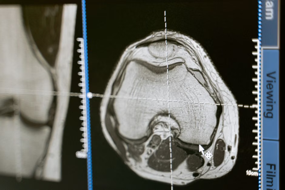
CT imaging uses X-rays to create detailed cross-sectional images of the brain. In stroke, it can show hyperdensities (bright areas) indicative of fresh blood in hemorrhagic stroke, or subtle signs of early edema and loss of gray-white matter differentiation in ischemic stroke. MRI, on the other hand, uses strong magnetic fields and radio waves to generate images. Advanced MRI sequences, like Diffusion-Weighted Imaging (DWI), are exquisitely sensitive to the restricted water movement that occurs within minutes of an ischemic stroke, making it a powerful tool for early detection.
The operational expertise we’ve gained since 1975, through developing over 25 instructional programs annually, shows us that understanding the fundamental principles of these technologies is vital for accurate interpretation. We teach that a non-contrast CT provides immediate answers regarding hemorrhage, while a CT angiogram (CTA) can visualize large vessel occlusions, crucial for thrombectomy decisions. MRI offers superior soft-tissue contrast, allowing for earlier detection of ischemic penumbra (at-risk tissue) and differentiating acute from chronic lesions, as discussed further in our programs on neuroradiology in clinical practice.
What To Look For: Signs and Symptoms of Acute Stroke
Recognizing the signs and symptoms of stroke is the first step toward rapid imaging. For healthcare professionals, a quick and systematic assessment using tools like the NIH Stroke Scale is essential. The symptoms often appear suddenly and can include:
- Sudden numbness or weakness on one side of the body, affecting the face, arm, or leg.
- Sudden confusion, trouble speaking, or difficulty understanding speech.
- Sudden trouble seeing in one or both eyes.
- Sudden trouble walking, dizziness, loss of balance, or lack of coordination.
- Sudden severe headache with no known cause.
Early identification of these symptoms should trigger immediate medical attention and expedited imaging protocols, as timely intervention significantly improves patient outcomes.
Ischemic Stroke MRI Findings: What Do We Look For?
For ischemic stroke, MRI, particularly Diffusion-Weighted Imaging (DWI), is incredibly sensitive. DWI detects cytotoxic edema within minutes of symptom onset, appearing as bright signal abnormalities. Combined with Apparent Diffusion Coefficient (ADC) maps that show corresponding low signal, this confirms acute ischemia. Perfusion-weighted imaging (PWI) helps identify the penumbra, the salvageable brain tissue at risk, guiding reperfusion therapy decisions.
In our practice, board-certified radiologists often combine DWI and PWI to calculate the “mismatch” – the difference between the irreversibly damaged core (DWI lesion) and the hypoperfused but potentially salvageable tissue (PWI lesion). This imaging biomarker is a key determinant for extended window thrombectomy in select patients. Additionally, Fluid-Attenuated Inversion Recovery (FLAIR) sequences can help estimate stroke onset time by looking for parenchymal hyperintensity, indicating that more than 4.5 hours have passed since symptom onset, a critical factor for thrombolysis eligibility.
CT vs MRI for Hemorrhagic Stroke: Key Distinctions
When it comes to hemorrhagic stroke, non-contrast CT is generally preferred for its speed and high sensitivity in detecting acute blood. Blood appears hyperdense (bright) on CT scans immediately after extravasation. While MRI can also detect hemorrhage, especially with specific sequences like susceptibility-weighted imaging (SWI), its initial role is typically secondary to CT due to longer acquisition times and potential patient contraindications.
Even though MRI can identify hemorrhage, particularly older hemorrhages or microbleeds, CT’s rapidity in the hyperacute setting is unmatched for ruling out intraparenchymal or subarachnoid hemorrhage. Since thrombolytic agents are contraindicated in hemorrhagic stroke, the ability of CT to rapidly and definitively exclude bleeding is a significant advantage in the initial emergency assessment.
“While MRI offers superior tissue characterization and is highly sensitive for acute ischemia, non-contrast CT remains the cornerstone for initial stroke imaging, primarily due to its rapid and accurate detection of intracranial hemorrhage, which is essential before administering thrombolytic therapy.”
Navigating Stroke Imaging Guidelines
Modern stroke imaging guidelines emphasize a tiered approach, starting with rapid CT to rule out hemorrhage, followed by more advanced imaging to characterize ischemic lesions and assess the penumbra. The goal is to identify patients who can benefit from reperfusion therapies as quickly and safely as possible.
Initial guidelines stress that a non-contrast head CT should be performed immediately upon presentation of stroke symptoms. If no hemorrhage is detected, a CT angiogram (CTA) and CT perfusion (CTP) may follow to evaluate for large vessel occlusion and penumbral mismatch, particularly in patients presenting within extended time windows. MRI is often used when the diagnosis remains unclear after CT, or in cases where posterior fossa stroke is suspected, which can be difficult to visualize on CT due to artifact. The decision tree for imaging is complex and constantly evolving, underscoring the need for continuous medical education.
Practical Tips for Optimizing Neuroimaging in Acute Stroke
For healthcare professionals, optimizing neuroimaging means making informed choices quickly. Here are some practical tips we emphasize in our accredited programs:
- Prioritize Speed: Always aim for the fastest possible imaging to exclude hemorrhage. Non-contrast CT is usually the quickest option available.
- Understand Modality Strengths: Recognize CT’s strength in detecting acute hemorrhage and bone abnormalities, versus MRI’s superiority for early ischemic changes and posterior fossa lesions.
- Know Your Local Protocols: Be familiar with your institution’s specific stroke protocols, including which imaging sequences are standard and the workflow for rapid interpretation.
- Consider Contraindications: Always screen patients for MRI contraindications (e.g., pacemakers, certain metallic implants) before ordering an MRI.
- Utilize Advanced Techniques: If available, use CTA and CTP with CT, or DWI/PWI with MRI, to identify large vessel occlusions and penumbral tissue for potential thrombectomy.
- Collaborate with Radiologists: Effective communication with the radiology team is paramount for appropriate imaging selection and accurate interpretation.
Navigating the complexities of neuroimaging in acute stroke demands not just technical proficiency but also a deep clinical understanding. Our collective experience, spanning over a century of combined expertise in CME program design and delivery, confirms that ongoing education empowers healthcare professionals to make the best decisions for their patients. Edu Symp, as an ACCME accredited provider, is committed to equipping you with the latest practical and evidence-based knowledge you need to excel. Visit edusymp.org to explore our wide range of accredited programs designed to enhance your clinical excellence and support lifelong learning.